We are thrilled to announce an esteemed panel of speakers who are at the forefront…
CurveBeam’s pedCAT Imaging System Exhibited at CIRMS Annual Meeting
The Council on Ionizing Radiation Measurements and Standards (CIRMS) 25th Annual Meeting in Gaithersburg, MD will showcase new cutting edge technologies touching on the fundamental aspects of radiation measurements and focusing on the theme of ‘Past, Present, and Future’. The dynamic and diverse aspects of the importance of measurements and standards in this area will be addressed by international experts from academia, industry, and government. These experts will examine radiation protection, industrial applications and radiation effects, medical applications, homeland security, and other related areas.
CurveBeam is pleased to announce we will be participating. A CurveBeam engineer will be presenting on Tuesday, March 28 during “Breakout Session III: Real Time Imaging for Orthopedic Applications.” Her talk is titled “Why Cone Beam CT Can Make 3D the Standard of Care in Extremity Imaging.” With Cone Beam CT imaging, CurveBeam is revolutionizing the way specialists diagnose and create comprehensive treatment plans for podiatric and orthopedic issues.
The core team behind CurveBeam pioneered Cone Beam CT imaging technology for the dental specialties. The introduction of point-of-care Cone Beam CT imaging revolutionized the industry and ushered in the advent of custom dental implants and improved practices in orthodontics and oral surgery. Today, Cone Beam CT scans are virtually the standard of care for advanced oral surgery treatment planning. In the same way, CurveBeam hopes to contribute to the improvement of the orthopedic and podiatric specialties worldwide through their new product pedCAT.
One of the goals of the CIRMS Annual Meeting is to get input from audience participants on the need for developing a 3D real-time imaging tool for evaluating orthotics inside shoes with the patient in a weight bearing position. Once the orthotic has been made, an image of the patient can be taken in the weight bearing position, with the patient standing in his/her shoes with the new orthotic installed in the shoe. This type of analysis could be used to evaluate if the orthotic does what it is meant to do. The 3D weight bearing images can provide information about specific bone alignment issues using the new orthotics. The images can also reveal if the new orthotic is providing the expected amount of correction without compromising other foot anatomical issues, and if the spacing between the major foot joints is affected by the new orthotic.
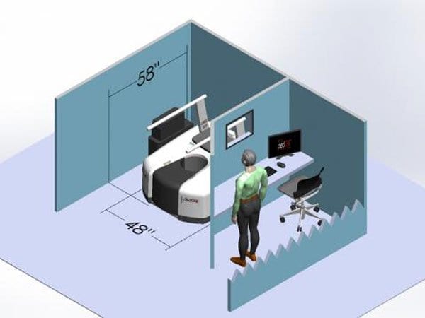
CurveBeams’s pedCAT system allows specialists a unique bilateral, weight bearing 3-dimensional view of the foot and ankle to fully diagnose and create comprehensive treatment plans. The pedCAT is a compact, ultra-low dose CT imaging system ideal for orthopedic and podiatric clinics. Patients benefit from the convenience of point-of-care advanced diagnostic imaging. Scan time is one minute, and the pedCAT automatically generates all standard X-Ray views in addition to the full CT volume. Depending on the scan protocol, the patient is exposed to 2 – 5 micro Sieverts per scan. That’s about the same as a plain X-Ray study of the foot and ankle, and less than the average daily background radiation a U.S. resident is exposed to.
.
To learn more about the CurveBeam pedCAT Imaging System and how it is revolutionizing the orthopedic and podiatric fields, visit curvebeamai.com, or talk with our team at the CIRMS 25th Annual Meeting, March 27th to March 29th in Gaithersburg.
Related Posts