Key Points: First tarsometatarsal joint (TMT1) hypermobility is associated with hallux valgus (HV) which is…
Examining the Added Value of Preoperative CT for Determining Cartilage Degeneration in Patients with Osteochondral Lesions of the Talar Dome
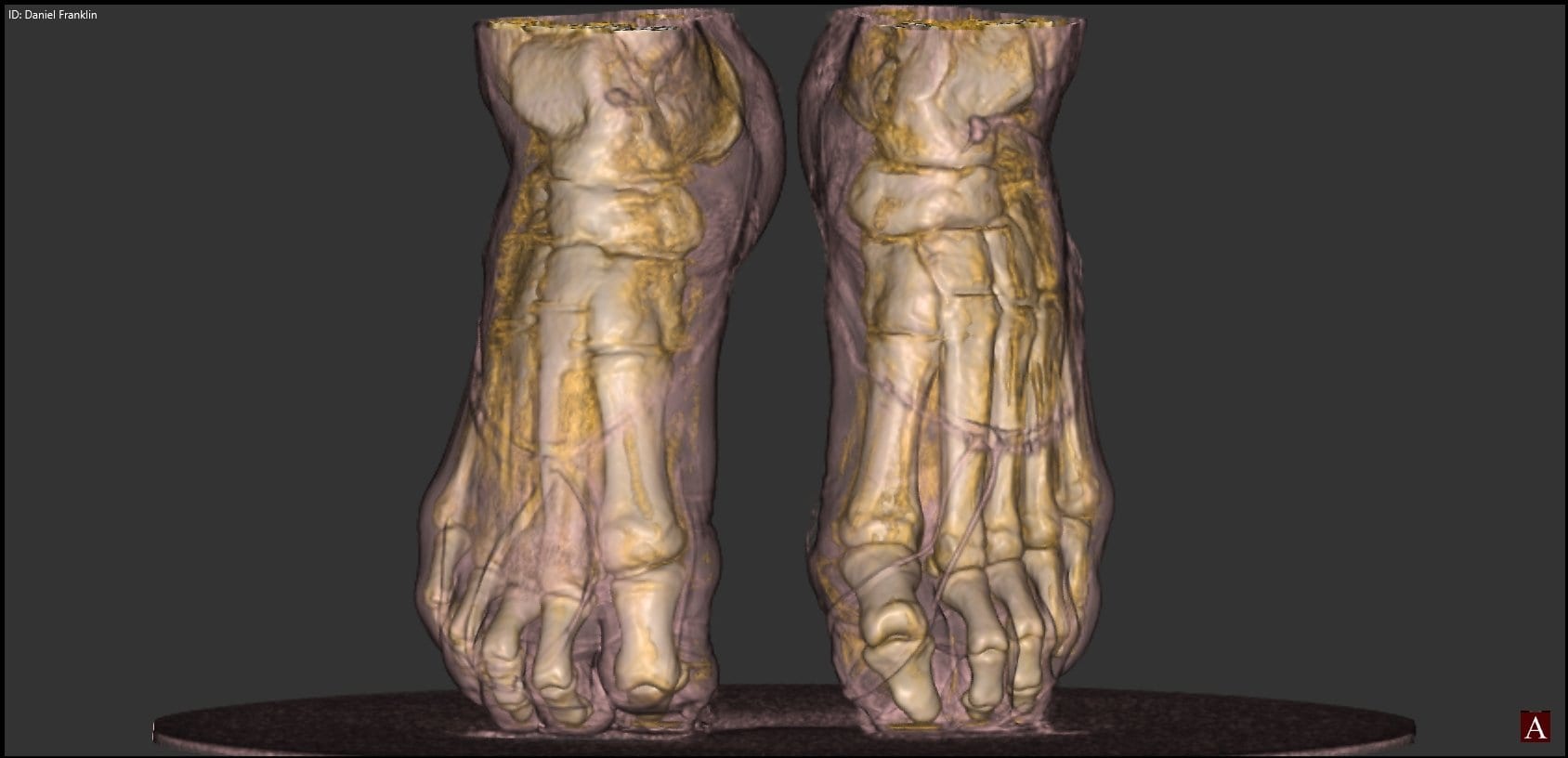
Osteochondral lesions of the talar dome (OLTs) involve the articular cartilage and subchondral bone. These lesions can cause deep ankle pain as well as impaired daily activities and sports activities. Surgical treatments are conducted as repair or replacement strategies to achieve biological healing of the OLT. The surgical procedure depends on the size, location, and stability of the lesion, as well as the extent of cartilage damage and the condition of the subchondral bone.
The usefulness of magnetic resonance imaging (MRI) has been reported to interpret the condition of the lesion ahead of a surgical procedure. It is not a perfect solution, however. An MRI might over- or underestimate the staging of OLT owing to bone edema and thinning of the articular cartilage. Computed tomography (CT) can provide more precise information than MRI on the subchondral bone, such as bone sclerosis, absorption, and cystic lesion.
To evaluate the unique features of the CT findings that relate to the condition of the articular cartilage in OLT, a study was conducted by the Department of Orthopaedic Surgery at Hiroshima University’s Graduate School of Biomedical Sciences in Hiroshima, Japan. The lead researcher on this study was Tomoyuki Nakasa, MD, PhD. The findings were published in the American Journal of Sports Medicine. Thirty ankles in 29 patients who had OLT with an osteochondral fragment were retrospectively reviewed to find out the extent to which CT image prediction of the histological findings on OLT will be useful to determine the most appropriate therapeutic strategy.
The osteochondral fragment of 19 ankles could be preserved by fixation or drilling at surgery. This was the preservation group. In the remaining 11 ankles, the osteochondral fragment was removed. This was the excision group. Preoperative CT findings were compared between the two groups to determine the relationship between the CT and histological findings. Biopsies of the osteochondral fragment from 13 ankles were also performed.
The area of lesion in the preservation group was significantly larger than that in the excision group. The CT images of the lesion showed the rate of absorption of the subchondral bone plate (SBP) in the preservation group to be lower than that in the excision group. The lesion bed absorption was higher in the preservation group than in the excision group. All cases in the excision group showed bed sclerosis, compared with 42.1% in the preservation group. The specimens with disruption of the SBP exhibited cartilage degeneration and abundant chondrocyte cloning. OLT with absorption of the SBP on CT showed severe cartilage degeneration, while the remaining SBP on CT showed low-grade cartilage degeneration.
In conclusion, while the condition of the SBP affects cartilage degeneration, CT findings provide important information for the determination of surgical treatment.
CurveBeam designs and manufactures Cone Beam CT imaging equipment for the orthopedic and podiatric specialties. Learn about CurveBeam’s Cone Beam CT systems here!
Related Posts