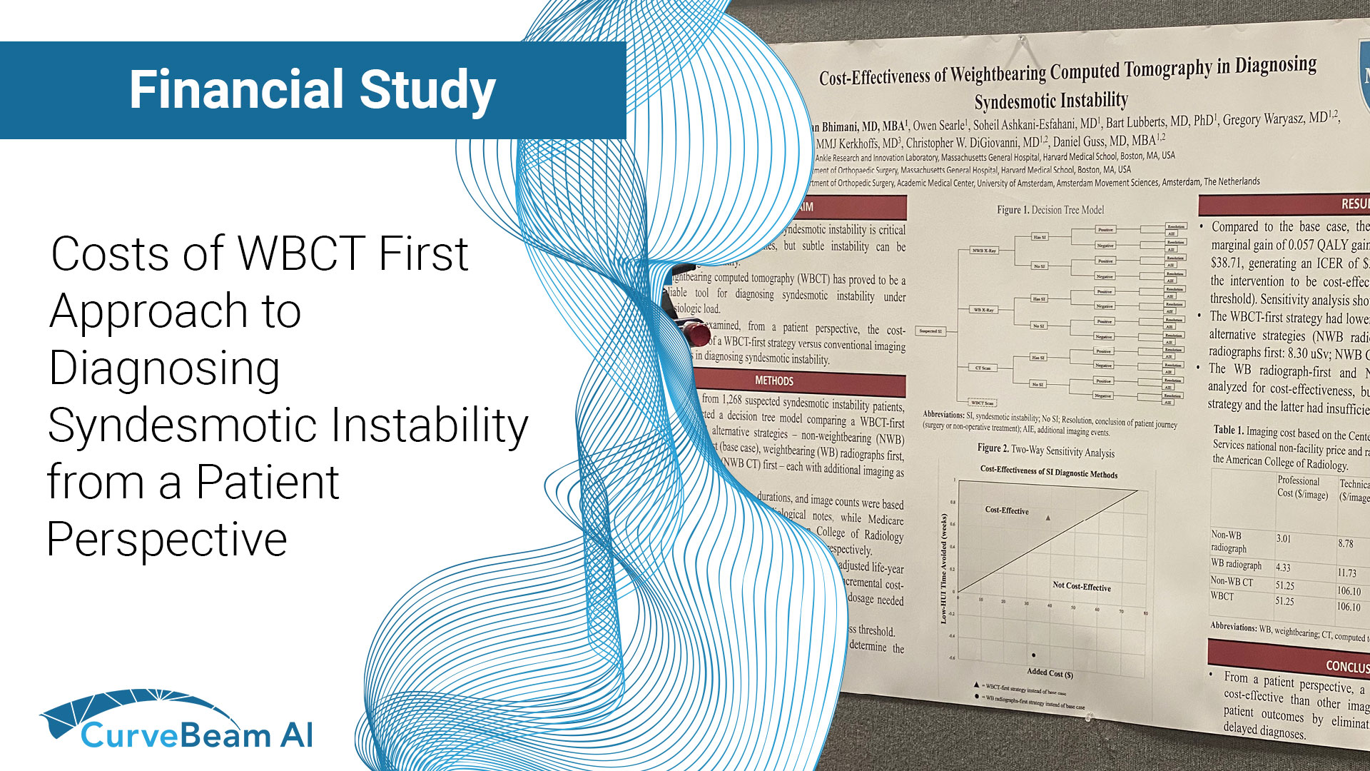
It is feasible for a single-practitioner podiatry practice to add weight bearing CT (WBCT) imaging and realize economical…
pedCAT: Weight Bearing CT Views of Lisfranc conditions
Lisfranc injuries can easily be missed on traditional radiographs.
One research study tried to determine the level of accuracy when using plain radiographs to assess midfoot conditions. Their results confirmed a high incidence of missed diagnosis even by experienced observers.¹
“Radiographic evaluation is crucial in the diagnosis…Weight bearing views are more sensitive since tarso-metatarsal instability may be revealed…The threshold for cross sectional imaging such as multi-detector CT should be low,” according to the study.
With the pedCAT, it is possible to get a weight bearing, three dimensional scan at the point of care. The tarso-metatarsal joint can be viewed without any superimposition from surrounding anatomy.
Delaying a 3D scan can be detrimental to recovery.
“The identification of Lisfranc joint injury can be difficult and is often not detected on initial presentation to the accident department. This is important because of the correlation between delay in treatment, particularly more than six months, and poor functional outcome,” the study reported.

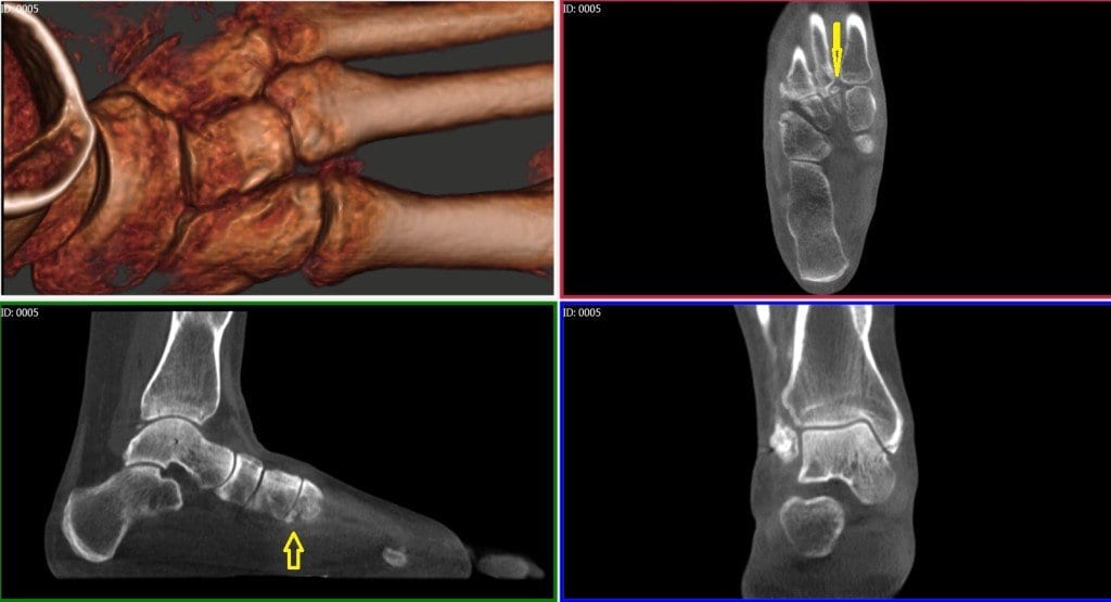
In this pedCAT scan, the 3D rendering shows the 2nd metatarsal is pushing on the 3rd metatarsal. The multi-planar slices reveal the fracture that is causing the dislocation. The sagittal and axial slices show a bone fragment as well. It would be impossible to see this information on a plain radiograph.

This pedCAT scan clearly depicts the gap between the first and second metatarsals.
¹ Sherief, Tamer. “Lisfranc injury: How frequently does it get missed? And how can we improve?” International Journal of the Care of the Injured. Elsevier Ltd. 10.106/j.injury.2006.10.002
Related Posts